
In collaborative work with Scott Murray, Bruno Olshausen, and David Woods, at the UC Davis Human Cognitive Neurophysiology Laboratory in the VA Medical Center, Martinez, California, and with Paul Schrater and the Center for Magnetic Resonance Imaging (CMRR) in Minnesota, we investigated the extent to which activity in primary visual cortex is influenced by high-level perceptual grouping (Murray et al., in press at PNAS). In one experiment, subjects viewed a perceptually bistable stimulus studied by Lorenceau and Shiffrar (1992). It consists of a line drawing of a diamond whose four corners are occluded by three vertical bars of the same color as the background.
Think of it as a diamond moving back and forth horizontally behind three invisible vertical bars (Panel A below). However, because the corners are hidden, sometimes the four segments appear to move up and down not as a single group, and sometimes they appear to move horizontally together as a single group. For a demonstration, try looking at the above quicktime movie of four horizontally moving line segments.
Can't see all four segments moving horizontally together? Then try this one for practice.
Still can't see the horizontal motion? OK, try this last version. There, you should see it.
Now go back to the original one and see if you can now see the four segments all moving together horizontally. We call the horizontal movement of all four segments, the coherent or diamond percept. This is when the shape is seen as a whole. When the four pieces don't appear move together, we call that an incoherent or non-diamond percept.
We measured activity in the primary visual cortex using BOLD functional Magnetic Resonance Imaging (fMRI). During the course of the experiment, the two percepts (coherent or incoherent) alternated after stable intervals of several to tens of seconds. FMRI measurements were taken while the subjects continuously viewed the stimulus and pressed buttons to indicate whether they were seeing the diamond or the non-diamond perceptual interpretations. Note that the visual stimulus was always the same--only the observer's perceptual interpretation changed.
Event-related averaging and cross correlation with a template, predicted from subjects' responses, were used to detect the BOLD response associated with each percept. Significant modulation of fMRI signals was detected in accordance with the percepts of the subjects. Significantly, the activity in V1 was less when a coherent diamond was perceived, and more when separate line segments were seen. Further, as shown below, the signal-to-noise ratio was sufficiently strong to reliably predict an individual observer's perceptual state on a trial-by-trial basis.
In panel B above, V1 activity (thin red lines) correlates with perceptual state (thick gray lines)--decreased activity is seen for the coherent percept. The signal in the time-series is sufficiently strong to reliably predict the perceptual state of the subject.
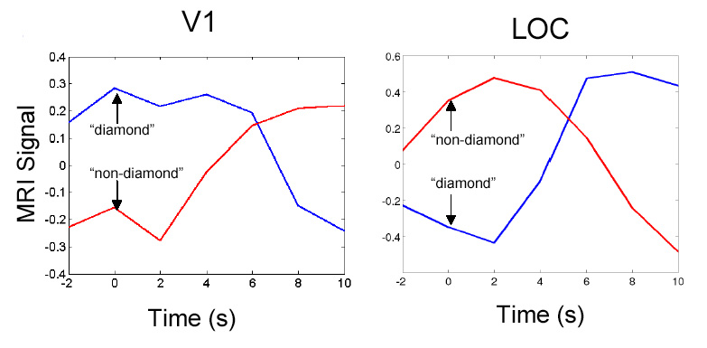
The above figures show an event-related average of the responses in V1 and LOC. You can see the dissociation of activity in the two areas and the perceptual states.
Activity level as early as the primary visual area can be modulated by high-level percepts in the absence of changes in the physical features of the visual stimuli. We argue that the sign of the modulation is consistent with the idea that neurons in these areas are engaged in predictive coding, perhaps through cortical back-projections.
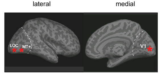
See the locations of V1 and LOC.
What would you predict for V1 responses when viewing moving random dots that cohere into a 3D shape vs. an incoherent set of motions? Or lines at random vs. organized into whole shapes? The answer is in the PNAS paper by Murray et al. (2002). Take a look at Scott Murray's description of the experiments.
References
Lorenceau, J. and M. Shiffrar (1992). "The influence of terminators on motion integration across space." Vision Res 32(2): 263-73.
Murray, S. O., D. Kersten, et al. (2002). "Shape perception reduces activity
in human primary visual cortex." Proc Natl Acad Sci U S A.
For a poster with figures, see: http://vision.psych.umn.edu/www/kersten-lab/papers/ARVO99fMRIPoster.pdf
Kersten Lab | Vision Lab | Psychology Department | University of Minnesota
© 2002 Computational Vision Lab, University of Minnesota, Department of
Psychology.